Monday, November 19, 2012

Observation #5 on November 13, 2012

    My 5th and final observation took place on November 13, 2012 from 3:45 p.m. until 5:52 p.m. with the assistance of Dr. McFarland in room 202 of the Hesler Biology Building on The University of Tennessee, Knoxville’s campus. The materials that were used during this observation were a Laborlux D (Leitz) microscope, a Sony Camera (HDR-HC9), a corresponding remote, and a Dell Computer. The microscope that I used this time was very difficult to work with because I constantly had to adjust the lense(s) after every movement. It had two Germany lenses (EF 10 and 3.5/0.10) and 1 exclusive Nikon lense. I removed the stand and lid from my MircroAquarium tank. Then I carefully laid the tank on the microscope stage with the open end away from me (Cook and McFarland 2013). The very first thing that I noticed was that there were barely any organisms left in my MicroAquarium.

   The first organism that I observed was an Actinosphaerium. There were at least 5-6 present throughout my 5th observation hidden near the mud. According to Dr. McFarland, they contain a big row of outer cells. Below is a picture of an Actinosphaerium that I captured during my 5th observation and edited with Adobe Photoshop Elements 7.0.


    The second organism that I observed was an Amoeba. This organism was found near the mud. Unlike in the previous observations, the Amoeba was not moving around much. It still showed flexibility but it was moving in one area very slowly. The foot was pushing organisms up very slowly. Below is an image of the Amoeba that I captured that was edited using Adobe Photoshop Elements 7.0.


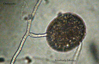
   The third organism that I observed during this 5th observation belonged to the phylum Oomycota. Dr. McFarland identified it as a type of water mold sporangia. It was very still and near the mud. It had hyphae connected to it. Moreover, Dr. McFarland dimmed the light on the camera and microscope to show the cytoplasmic movement inside of the hyphae. I saw 2 of these organisms.


   The fourth organism that I observed was an Arcella and I only found one near the mud.
 
 
   The next organism that I observed was a Rotifer. Dr. Mcfarland explained that Rotifers are like vacuum cleaners because they do not eat the plants, however they do eat what's on the plants. They eat very fast. The way that they look reminds me of rats. Rotifers move very fast and they do what seems to be flips. I saw 3 of these organisms during my 5th observation.


   The next organism I observed was a Colops. Dr. McFarland also stated that it was a ciliate. It moved fairly quickly. This organism has long cilia (hairs) behind it. I only saw one of its kind.


   The final organism that I observed during my 5th and final observation was a Cyclops. Dr. McFarland explained that the red spot that is on it helps it sense light. This was the only Cyclops I saw during this observation.


   In conclusion, I disliked this observation the most out of all because there were not a lot of new organisms like in my previous observations. It was very boring. I saw one diatom that I did not get a chance to snap a picture of. Also, I saw worm castings present throughout and a few little flagelletes throughout. At the end of this observation instead of replacing the water that was lost,  Dr. McFarland instructed me to place my MicroAquarium in the black tray located by the entrance because I was done with my final observation. Hooray!
 
 
References:
 
 
Cook R, McFarland K. 2013. General Botany 111 Laboratory Manual. 14th ed, Knoxville (TN).155-157 p.

Citation: (Cook and McFarland 2013)


Monday, November 12, 2012

Observations #4 11/7/12

   
     On November 7, 2012 from about 3:35 pm - 5:30 pm is when my fourth observation took place. As usual, my observation took place with the assistance of Dr. McFarland in room 202 of the Hesler Biology Building on The University of Tennessee, Knoxville’s campus. The materials that were used during this observation were a CH30 Olympus microscope, a Sony Camera (HDR-HC9), a corresponding remote, and a Dell Computer. As a reminder, the water in my MicroAquarium is from source 10 which was taken from a water pool below spring; Lynnhurst Cemetery (McFarland 2012). To begin observing I removed the stand and lid from my MircroAquarium tank. Then I carefully laid the tank on the microscope stage with the open end away from me (Cook and McFarland 2013). It became very clear that all of my large amounts of big organisms from last week were gone. However, most organisms appeared to be near the bottom.


     According to Dr. McFarland, the first organism that I observed was an Amoeba. It was clear and stretching with so many organisms visible on the inside of it. The Amoeba was very flexible. The foot of it was pushing things to the top of it. Dr. McFarland classified its movement as being cytoplasmic and he explained to me that there are more Amoebas in a pond than there are of us humans on Earth. Also there were a few nuclides visible that were on the inside of the organism. Throughout my observation there were so many of these Amoebas found everywhere in my MicroAquarium. I found at least 6 although I am quite sure that there were many more.

Below is a picture of an Amoeba from my MicroAquarium.


       The next organism I observed was an Actinosphaerium (Hedley and Patterson 1996). It appeared very still and had little to no movement. There were many ciliates floating around it. I observed so many of these organisms, it was so hard to keep count. There were well over 7 found during this observation. Most of the Actinosphaeriums were found along the bottom of my MicroAquarium near the mud. Dr. McFarland explained that these organisms have a big ball on their side which is a vacuole that pops and disappears. During this observation, he took an extended approach and decided that we should measure the size of a big Actinosphaerium and a smaller one. So, by using the microscope Dr. McFarland came to the conclusion that under the 10x and 40x lense the bigger Actinosphaerium was 0.14 micrometers (57 units using 40x and 14 units using 10x) and the smaller one was 0.02 micrometers (.8 units using 40x and 2 units using 10x).

Below is a comparison using the 10x lense of a big Actinosphaerium and a smaller one. If you look closely at the image below the vacuole on the small Actinosphaerium is visible.


      The next organism the I observed was a Rotifer. I seen about 2 throughout my observation on this week. It was hanging on to the Utricularia gibba plant by its tail (McFarland 2012). This is the carnivous and flowering plant that was placed in the MicroAquarium during the initial setup (McFarland 2012).

Below is a picture of the Rotifer that I observed during my observation.


      The next organism I observed was very unique and Dr. McFarland identified it as chained Spirillum bacteria of something small although the scientific name was not evident. This was the only pair of the organism(s) that I seen and there was little movement between the organism(s). 
 
 
Below is an image of the chained bacteria Spirillum.


       The final organism that I observed during the week was a Halteria. It was located near the bottom. I only seen one of this organism although I am sure that there were many more. It was very hard to capture an image of it because it moves so quick. According to Dr. McFarland, under the 40x lense it was about 10 units and 0.24 micrometers.
 
Below is an image of the Halteria. 


     Throughout my observation(s) on this week I observed a few other organisms (Midge, flagella, and a water flea) that I was unable to capture pictures of due to their fast movements. The Midge appeared to be eating on the Amblestegium sp. which is from the moss collection (McFarland 2012). According to Dr. McFarland, it belongs to the Phylum Arthropoda. Also the water flea was down near the mud and had a red dot on it which helped Dr. McFarland identify it. It moved extremely quick. Moreover, I also observed some worm castings near the mud. There was also some flagella that were very small and each possessed a tail trailing it. Dr. McFarland stated that they eat small particles. 
 
 
     All images from this observation were edited using Adobe Photoshop Elements 7.0. In addition, at the end of my observation(s) to replace the water that may have been lost in my MicroAquarium  somehow, I added more to it with the clear water bottle that Dr. McFarland provided.
 
References (CBE Style):
 
Cook R, McFarland K. 2013. General Botany 111 Laboratory Manual. 14th ed, Knoxville (TN).155-157 p.

Citation: (Cook and McFarland 2013)

McFarland K. Botany 111 Fall 2012. [Internet]. 2012. Available from: http://botany1112012.blogspot.com/

Citation: (McFarland 2012)
 Hedley S and Patterson D. 1996. Free-Living Freshwater Protozoa "A Colour Guide". Corringham Road, London: Manson Publishing. 169 p. Figure 394.

Citation: (Hedley and Patterson 1996)


Monday, November 5, 2012

Observation #3 October 31, 2012

Observation #3 on October 31, 2012
            On October 26, 2012 Dr. McFarland inserted one Beta Food Pellet into my MicroAquarium. My observation (s) took place on October 31, 2012 at approximately 2:15 p.m. My observation (s) took place in the usual room 202 of the Hesler Biology Building on The University of Tennessee, Knoxville’s campus with the help of Dr. McFarland. Just like in my last observation, I used a CH30 Olympus microscope, a Sony Camera (HDR-HC9), and a Dell Computer. I also used the corresponding remote to snap images of interesting organisms that I found throughout my observation To observe I removed the stand and lid from the MicroAquarium tank. Then I carefully laid the tank on the microscope stage with the open end away from me.  Since October 23, 2012 (my last observation) and with the Beta Food Pellet added on October 26th, there were a lot more organisms that were visible than there were in the previous observations. To help identify organisms Dr. McFarland and I used the book, An Illustrated Guide to the Protozoa.

            The first organism that I observed is related to the Amoebas. I observed this using a Nikon Japan lense (40). Dr. McFarland helped me identify that it was of the Genus Microcorycia (Bovee, Lee, Lynn, and Small 1985). This organism possesses a finger like structure that pops out. However, during my observation (s) the organism did not pop the structure out.  It was very still. This was the only one of the organism that I seen while observing. Below is an image of the organism edited with Adobe Photoshop Elements 7.0.


            The second organism that I observed was a Stentor sp. (Bovee, Lee, Lynn, and Small 1985). To me, it looks like a trumpet. Individual cilia were pushing water into its funnel during my observation(s) of it. I observed and captured a picture of it using the Nikon lense. Below is an image of the Stentor organism edited using Photoshop. I combined two shots of the organism the top picture shows it upclose under the Nikon lense and the bottom image attached shows the Stentor sp. under the EF 10 (1.25) lense.


            The third organism that I observed was a Halteria sp. (Bovee, Lee, Lynn, and Small 1985). There were a lot of them. The organism moved so quickly. On the outer surface of the Halteria there were cilia flowing. I saw the specific organism that I captured a photo of towards the bottom near the mud.


            The fourth organism that I observed was a Litonotus sp. (Bovee, Lee, Lynn, and Small 1985). Notice in the image below the vacuole on the back end. According to Dr. McFarland, it is a contractual vacuole that gets rid of excess water in the cell. This organism moved very quickly and I seen about 3 or 4 throughout my observation (s).


            The fifth organism that I observed was very still. It is an Arcella sp. (Bovee, Lee, Lynn, and Small 1985). It just layed and kept very stiff in the middle of nowhere.


            The final organism that I observed was an Amoeba Radiosa sp. (Bovee, Lee, Lynn, and Small 1985). This organism catches its observer’s eye as you can see in the image below. While I was observing this organism it moved at medium speed in the middle of the aquarium. Dr. McFarland said during my observation(s), “It can become one of your worst nightmares at night if you think about it too long.”


            Overall, this was my favorite observation out of all because I found a lot of organisms. I’m sure there were more, but the Genus Microcorycia, Stentor sp., Halteria sp., Litonotus sp., Arcella sp., and the Amoeba Radiosa sp were the ones that Dr. McFarland and I received the chance to capture. Like instructed on my last observation(s), at the end of my observation(s) to replace the water that may have been lost somehow, I added more to it with the clear water bottle that he provided.


References (CBE Style):

 Bovee E, Lee J, Lynn D, Small E. 1985. An Illustrated Guide to the Protozoa. 80 p., 91 p., 107 p., 132 p., 139p.

 Citation: (Bovee, Lee, Lynn, and Small 1985)

 
Cook R, McFarland K. 2013. General Botany 111 Laboratory Manual. 14th ed, Knoxville (TN).155-157 p.


Citation: (Cook and McFarland 2013)
 
McFarland K. Botany 111 Fall 2012. [Internet]. 2012. Available from: http://botany1112012.blogspot.com/


Citation: (McFarland 2012)


Monday, October 29, 2012

Observation #2 10/23/12


Below is an image of my MicroAquarium that I took before I started my second Observation using my LG Beacon phone.


My second observation occurred on October 23, 2012 at approximately 5 o clock p.m. with the assistance of Dr. McFarland in room 202 of the Hesler Biology Building on The University of Tennessee, Knoxville’s campus. It lasted until about 6:15 p.m. The materials that were used during this observation were a CH30 Olympus microscope, a Sony Camera (HDR-HC9), and a Dell Computer. The Sony camera was hooked up to the microscope and was used to project the images through the Dell computer screen. A corresponding remote was also used to snap images of interesting organisms that I found throughout my observation. Moreover, the microscope contained an EA 4 lense, an EA 10 lense, and a very special lense which was an exclusive Nikon Japan lense (.55LWD). To observe I removed the stand and lid from the MircroAquarium tank. Then I carefully laid the tank on the microscope stage with the open end away from me. I first observed some little organisms that had cilia hair-like organelles which Dr. McFarland later told me were only ciliates. The ciliates were moving throughout the aquarium in no specific order. The next organism that I observed was an Actinosphaerium (Hedley and Patterson 1996).   It is a multinucleated heliozoon and is related to Actinophrys (Hedley and Patterson 1996). It appeared to be floating throughout. The final organism that I observed on that day was an Oscillatoria Splendida (Forest 1954). This organism had a photosynthetic bacterial sheath around it, according to Dr. McFarland. It was sticking out from the sample of the Amblestegium (from the moss collection) plant. Under the EA 4 lense of the microscope, the worm-like cyanobacteria photosynthetic organism appeared small. Moreover, with the EA 10 lense the worm appeared skinny and very long. Once I used the exclusive Japan lense I captured a good image of it because it projected the organism the greatest and produced the best image out all three lenses. To create or perfect the images below, Dr. McFarland showed me how to use Photoshop to crop and make the images appear much better.

Finally, at the end of my observation before I placed my MicroAquarium back in its appropriate section, Dr. McFarland instructed me to replace the water that may have been lost somehow, by adding more to it with the clear water bottle that he provided. Overall, I observed more organisms last time, but the two organisms that I did observe this time were very original and new to my eyes; although there was only one of each of the two organisms besides the ciliates this time.


Below is an image of the Actinosphaerium that I edited under the assistance of Dr. McFarland using Photoshop.


Below is an image of the Oscillatoria Splendida that I edited under the assistance of Dr. McFarland using Photoshop.


References:

Cook R, McFarland K. 2013. General Botany 111 Laboratory Manual. 14th ed, Knoxville (TN).155-157 p.

Citation: (Cook and McFarland 2013)

Forest H.1954. Handbook of Algae. The University of Tennessee Press. 387 p. Figure 13.

Citation: (Forest 1954)

Hedley S and Patterson D. 1996. Free-Living Freshwater Protozoa “A Colour Guide”. Corringham Road, London: Manson Publishing. 169 p. Figure 394.

Citation: (Hedley and Patterson 1996)


Monday, October 22, 2012

Observation Date: October 16, 2012


Observation Date: October 16, 2012
I began setting up my MicroAquarium on October 16, 2012 with the assistance of Ms. Massana and Dr. McFarland. In setting up my MicroAquarium, my water source was from source 10 which was taken from a water pool below spring; Lynnhurst Cemetery (McFarland 2012). The cemetery is located off of Adair Drive (McFarland 2012). It is located in Knox Co; Knoxville, Tennessee (McFarland 2012). The water is exposed to partial shade (McFarland 2012). It is the Spring Feed Pond (N36 01.357 W83 55.731 958 ft.). The water was collected by Dr. McFarland on October 9, 2011 (McFarland 2012).

            Meanwhile to set up my MicroAquarium I first made the decision to choose source 10 as my water source. Then, I obtained a MicroAquarium which was basically a glass tank, a stand holder, and a lid (Cook and McFarland 2013). I color coordinated it with three dots respectively to represent my lab section which is 001 (green), my table 6 (orange),and my seat # which was 4. Next, I recorded my initials in order with one letter being placed on each of the three dots (KSM). Then, I gathered a pipet and extracted water from source 10. When gathering water, I first sucked up enough mud from the source then I gradually began to get water(from the closest to the mud to the surface of the water). I then placed a sample of the plant Amblestegium and a sample of the plant Utricularia gibba into my MicroAquarium. Amblestegium (from the moss collection) was collected from Natural spring at Carters Mill Park, Carter Mill Road, Knox Co. TN on October 9, 2011 by Dr. McFarland (McFarland 2012). It endured some partial shade exposure (McFarland 2012). Utricularia gibba is both a flowering and carnivorous plant it was also collected on October 9, 2011 by Dr. McFarland from south shore of Spain Lake (N 35o55 12.35" W088o20' 47.00), Camp Bella Air Rd. East of Sparta Tn. in White Co (McFarland 2012). However, since then it has been grown in water tanks outside of greenhouse at Hesler Biology Building. The University of Tennessee. Knox Co. Knoxville TN (McFarland 2012).

            Once I set up my MicroAquarium I then took the initiative to examine it and its contents under a microscope. I used the 4x and 10x lens, but I began by using the 4x lens to get a good focus. I did not use the 40x lens because it would have caused my MicroAquarium to break. To observe I removed the stand and lid from the MircroAquarium tank. Then I carefully laid the tank on the microscope stage with the open end away from me. I was amazed that the water didn’t come out which Dr. McFarland explained was due to surface tension. Once under the microscope, I saw moving organisms more specifically a big brown beetle shaped insect and a really small circular insect that was floating throughout the top and middle of the aquarium. Another organism that I observed on October 16, 2012 had a heart-shaped back and 4 legs with 5 extensions from each leg and appeared to float throughout the top and middle of the aquarium. There was another organism at the very bottom that looked as if it were eating on the Amblestegium plant. I only seen one of each of the organisms that I described. I am looking forward to the next obeservations of my MicroAquarium.

References (CBE Style)

Cook R, McFarland K. 2013. General Botany 111 Laboratory Manual. 14th ed, Knoxville (TN).155-157 p.

Citation: (Cook and McFarland 2013)

McFarland K. Botany 111 Fall 2012. [Internet]. 2012. Available from : http://botany1112012.blogspot.com/

CItation: (McFarland 2012)