The first organism that I observed was an Actinosphaerium. There were at least 5-6 present throughout my 5th observation hidden near the mud. According to Dr. McFarland, they contain a big row of outer cells. Below is a picture of an Actinosphaerium that I captured during my 5th observation and edited with Adobe Photoshop Elements 7.0.
The second organism that I observed was an Amoeba. This organism was found near the mud. Unlike in the previous observations, the Amoeba was not moving around much. It still showed flexibility but it was moving in one area very slowly. The foot was pushing organisms up very slowly. Below is an image of the Amoeba that I captured that was edited using Adobe Photoshop Elements 7.0.
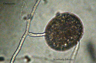
The third organism that I observed during this 5th observation belonged to the phylum Oomycota. Dr. McFarland identified it as a type of water mold sporangia. It was very still and near the mud. It had hyphae connected to it. Moreover, Dr. McFarland dimmed the light on the camera and microscope to show the cytoplasmic movement inside of the hyphae. I saw 2 of these organisms.
The fourth organism that I observed was an Arcella and I only found one near the mud.
The next organism that I observed was a Rotifer. Dr. Mcfarland explained that Rotifers are like vacuum cleaners because they do not eat the plants, however they do eat what's on the plants. They eat very fast. The way that they look reminds me of rats. Rotifers move very fast and they do what seems to be flips. I saw 3 of these organisms during my 5th observation.
The next organism I observed was a Colops. Dr. McFarland also stated that it was a ciliate. It moved fairly quickly. This organism has long cilia (hairs) behind it. I only saw one of its kind.
The final organism that I observed during my 5th and final observation was a Cyclops. Dr. McFarland explained that the red spot that is on it helps it sense light. This was the only Cyclops I saw during this observation.
In conclusion, I disliked this observation the most out of all because there were not a lot of new organisms like in my previous observations. It was very boring. I saw one diatom that I did not get a chance to snap a picture of. Also, I saw worm castings present throughout and a few little flagelletes throughout. At the end of this observation instead of replacing the water that was lost, Dr. McFarland instructed me to place my MicroAquarium in the black tray located by the entrance because I was done with my final observation. Hooray!
References:
Cook R,
McFarland K. 2013. General Botany 111 Laboratory Manual. 14th ed, Knoxville
(TN).155-157 p.
Citation: (Cook and McFarland 2013)


